THE EVOLUTION OF MAN
Volume II
CHAPTER XX
OUR WORM-LIKE ANCESTORS
The gastræa theory has now convinced us that all the Metazoa or multicellular animals can be traced to a common stem-form, the Gastræa. In accordance with the biogenetic law, we find solid proof of this in the fact that the two-layered embryos of all the Metazoa can be reduced to a primitive common type, the gastrula. Just as the countless species of the Metazoa do actually develop in the individual from the simple embryonic form of the gastrula, so they have all descended in past time from the common stem-form of the Gastræa. In this fact, and the fact we have already established that the Gastræa has been evolved from the hollow vesicle of the one-layered Blastæa, and this again from the original unicellular stem-form, we have obtained a solid basis for our study of evolution. The clear path from the stem-cell to the gastrula represents the first section of our human stem-history (Chapters VIII, IX, and XIX).
The second section, that leads from the Gastræa to the Prochordonia, is much more difficult and obscure. By the Prochordonia we mean the ancient and long-extinct animals which the important embryonic form of the chordula proves to have once existed (cf. Figs. 83–86). The nearest of living animals to this embryonic structure are the lowest Tunicates, the Copelata (Appendicaria) and the larvæ of the Ascidia. As both the Tunicates and the Vertebrates develop from the same chordula, we may infer that there was a corresponding common ancestor of both stems. We may call this the Chordæa, and the corresponding stem-group the Prochordonia or Prochordata.
From this important stem-group of the unarticulated Prochordonia (or “primitive chorda-animals”) the stems of the Tunicates and Vertebrates have been divergently evolved. We shall see presently how this conclusion is justified in the present condition of morphological science.
We have first to answer the difficult and much-discussed question of the development of the Chordæa from the Gastræa; in other words, “How and by what transformations were the characteristic animals, resembling the embryonic chordula, which we regard as the common stem-forms of all the Chordonia, both
[ 219 ]
Tunicates and Vertebrates, evolved from the simplest two-layered Metazoa?”
The descent of the Vertebrates from the Articulates has been maintained by a number of zoologists during the last thirty years with more zeal than discernment; and, as a vast amount has been written on the subject, we must deal with it to some extent. All three classes of Articulates in succession have been awarded the honour of being considered the “real ancestors” of the Vertebrates: first, the Annelids (earth-worms, leeches, and the like), then the Crustacea (crabs, etc.), and, finally, the Tracheata (spiders, insects, etc.). The most popular of these hypotheses was the annelid theory, which derived the Vertebrates from the Worms. It was almost simultaneously (1875) formulated by Carl Semper, of Würtzburg, and Anton Dohrn, of Naples. The latter advanced this theory originally in favour of the failing degeneration theory, with which I dealt in my work, Aims and Methods of Modern Embryology.
This interesting degeneration theory—much discussed at that time, but almost forgotten now—was formed in 1875 with the aim of harmonising the results of evolution and ever-advancing Darwinism with religious belief. The spirited struggle that Darwin had occasioned by the reformation of the theory of descent in 1859, and that lasted for a decade with varying fortunes in every branch of biology, was drawing to a close in 1870–1872, and soon ended in the complete victory of transformism. To most of the disputants the chief point was not the general question of evolution, but the particular one of “man’s place in nature”—“the question of questions,” as Huxley rightly called it. It was soon evident to every clear-headed thinker that this question could only be answered in the sense of our anthropogeny, by admitting that man had descended from a long series of Vertebrates by gradual modification and improvement.
In this way the real affinity of man and the Vertebrates came to be admitted on all hands. Comparative anatomy and ontogeny spoke too clearly for their testimony to be ignored any longer. But in order still to save man’s unique position, and especially the dogma of personal immortality, a number of natural philosophers and theologians discovered an admirable way of escape in the “theory of degeneration.” Granting the affinity, they turned the whole evolutionary theory upside down, and boldly contended that “man is not the most highly developed animal, but the animals are degenerate men.” It is true that man is closely related to the ape, and belongs to the vertebrate stem; but the chain of his ancestry goes upward instead of downward. In the beginning “God created man in his own image,” as the prototype of the perfect vertebrate; but, in consequence of original sin, the human race sank so low that the apes branched off from it, and afterwards the lower Vertebrates. When this theory of degeneration was consistently developed, its supporters were bound to hold that the entire animal kingdom was descended from the debased children of men.
This theory was most strenuously defended by the Catholic priest and natural philosopher, Michelis, in his Hæckelogony: An Academic Protest against Hæckel’s Anthropogeny (1875). In still more “academic” and somewhat mystic form the theory was advanced by a natural philosopher of the older Jena school—the mathematician and physicist, Carl Snell. But it received its chief support on the zoological side from Anton Dohrn, who maintained the anthropocentric ideas of Snell with particular ability. The Amphioxus, which modern science now almost unanimously regards as the real Primitive Vertebrate, the ancient model of the original vertebrate structure, is, according to Dohrn, a late, degenerate descendant of the stem, the “prodigal son” of the vertebrate family. It has descended from the Cyclostoma by a profound degeneration, and these in turn from the fishes; even the Ascidia and the whole of the Tunicates are merely degenerate fishes! Following out this curious theory, Dohrn came to contest the general belief that the Cœlenterata and Worms are “lower animals”; he even declared that the unicellular Protozoa were degenerate Cœlenterata. In his opinion “degeneration is the great principle that explains the existence of all the lower forms.”
If this Michelis-Dohrn theory were true, and all animals were really degenerate descendants of an originally perfect humanity, man would assuredly be the true centre and goal of all terrestrial life; his anthropocentric position and his immortality would be saved. Unfortunately, this trustful theory is in such
[ 220 ]
flagrant contradiction to all the known facts of paleontology and embryology that it is no longer worth serious scientific consideration.
But the case is no better for the much-discussed descent of the Vertebrates from the Annelids, which Dohrn afterwards maintained with great zeal. Of late years this hypothesis, which raised so much dust and controversy, has been entirely abandoned by most competent zoologists, even those who once supported it. Its chief supporter, Dohrn, admitted in 1890 that it is “dead and buried,” and made a blushing retraction at the end of his Studies of the Early History of the Vertebrate.
Now that the annelid-hypothesis is “dead and buried,” and other attempts to derive the Vertebrates from Medusæ, Echinoderms, or Molluscs, have been equally unsuccessful, there is only one hypothesis left to answer the question of the origin of the Vertebrates—the hypothesis that I advanced thirty-six years ago and called the “chordonia-hypothesis.” In view of its sound establishment and its profound significance, it may very well claim to be a theory, and so should be described as the chordonia or chordæa theory.
I first advanced this theory in a series of university lectures in 1867, from which the History of Creation was composed. In the first edition of this work (1868) I endeavoured to prove, on the strength of Kowalevsky’s epoch-making discoveries, that “of all the animals known to us the Tunicates are undoubtedly the nearest blood-relatives of the Vertebrates; they are the most closely related to the Vermalia, from which the Vertebrates have been evolved. Naturally, I do not mean that the Vertebrates have descended from the Tunicates, but that the two groups have sprung from a common root. It is clear that the real Vertebrates (primarily the Acrania) were evolved in very early times from a group of Worms, from which the degenerate Tunicates also descended in another and retrogressive direction.” This common extinct stem-group are the Prochordonia; we still have a silhouette of them in the chordula-embryo of the Vertebrates and Tunicates; and they still exist independently, in very modified form, in the class of the Copelata (Appendicaria, Fig. 225).
The chordæa-theory received the most valuable and competent support from Carl Gegenbaur. This able comparative morphologist defended it in 1870, in the second edition of his Elements of Comparative Anatomy; at the same time he drew attention to the important relations of the Tunicates to a curious worm, Balanoglossus: he rightly regards this as the representative of a special class of worms, which he called “gut-breathers” (Enteropneusta). Gegenbaur referred on many other occasions to the close blood-relationship of the Tunicates and Vertebrates, and luminously explained the reasons that justify us in framing the hypothesis of the descent of the two stems from a common ancestor, an unsegmented worm-like animal with an axial chorda between the dorsal nerve-tube and the ventral gut-tube.
The theory afterwards received a good deal of support from the research made by a number of distinguished zoologists and anatomists, especially C. Kupffer, B. Hatschek, F. Balfour, E. Van Beneden, and Julin. Since Hatschek’s Studies of the Development of the Amphioxus gave us full information as to the embryology of this lowest vertebrate, it has become so important for our purpose that we must consider it a document of the first rank for answering the question we are dealing with.
The ontogenetic facts that we gather from this sole survivor of the Acrania are the more valuable for phylogenetic purposes, as paleontology, unfortunately, throws no light whatever on the origin of the Vertebrates. Their invertebrate ancestors were soft organisms without skeleton, and thus incapable of fossilisation, as is still the case with the lowest vertebrates—the Acrania and Cyclostoma. The same applies to the greater part of the Vermalia or worm-like animals, the various classes and orders of which differ so much in structure. The isolated groups of this rich stem are living branches of a huge tree, the greater part of which has long been dead, and we have no fossil evidence as to its earlier form. Nevertheless, some of the surviving groups are very instructive, and give us clear indications of the way in which the Chordonia were developed from the Vermalia, and these from the Cœlenteria.
While we seek the most important of these palingenetic forms among the groups of Cœlenteria and Vermalia, it is understood that not a single one of them
[ 221 ]
must be regarded as an unchanged, or even little changed, copy of the extinct stem-form. One group has retained one feature, another a different feature, of the original organisation, and other organs have been further developed and characteristically modified. Hence here, more than in any other part of our genealogical tree, we have to keep before our mind the full picture of development, and separate the unessential secondary phenomena from the essential and primary. It will be useful first to point out the chief advances in organisation by which the simple Gastræa gradually became the more developed Chordæa.
We find our first solid datum in the gastrula of the Amphioxus (Figure 1.38). Its bilateral and tri-axial type indicates that the Gastræads—the common ancestors of all the Metazoa—divided at an early stage into two divergent groups. The uni-axial Gastræa became sessile, and gave rise to two stems, the Sponges and the Cnidaria (the latter all reducible to simple polyps like the hydra). But the tri-axial Gastræa assumed a certain pose or direction of the body on account of its swimming or creeping movement, and in order to sustain this it was a great advantage to share the burden equally between the two halves of the body (right and left). Thus arose the typical bilateral form, which has three axes. The same bilateral type is found in all our artificial means of locomotion—carts, ships, etc.; it is by far the best for the movement of the body in a certain direction and steady position. Hence natural selection early developed this bilateral type in a section of the Gastræads, and thus produced the stem-forms of all the bilateral animals.
The Gastræa bilateralis, of which we may conceive the bilateral gastrula of the amphioxus to be a palingenetic reproduction, represented the two-sided organism of the earliest Metazoa in its simplest form. The vegetal entoderm that lined their simple gut-cavity served for nutrition; the ciliated ectoderm that formed the external skin attended to locomotion and sensation; finally, the two primitive mesodermic cells, that lay to the right and left at the ventral border of the primitive mouth, were sexual cells, and effected reproduction. In order to understand the further development of the gastræa, we must pay particular attention to: (1) the careful study of the embryonic stages of the amphioxus that lie between the gastrula and the chordula; (2) the morphological study of the simplest Platodes (Platodaria and Turbellaria) and several groups of unarticulated Vermalia (Gastrotricha, Nemertina, Enteropneusta).
We have to consider the Platodes first, because they are on the border between the two principal groups of the Metazoa, the Cœlenteria and the Cœlomaria. With the former they share the lack of body-cavity, anus, and vascular system; with the latter they have in common the bilateral type, the possession of a pair of nephridia or renal canals, and the formation of a vertical brain or cerebral ganglion. It is now usual to distinguish four classes of Platodes: the two free-living classes of the primitive worms (Platodaria) and the coiled-worms (Turbellaria), and the two parasitic classes of the suctorial worms (Trematoda) and the tape-worms (Cestoda). We have only to consider the first two of these classes; the other two are parasites, and have descended from the former by adaptation to parasitic habits and consequent degeneration.
The primitive worms (Platodaria) are very small flat worms of simple construction, but of great morphological and phylogenetic interest. They have been hitherto, as a rule, regarded as a special order of the Turbellaria, and associated with the Rhabdocœla; but they differ considerably from these and all the other Platodes (flat worms) in the absence of renal canals and a special central nervous system; the structure of their tissue is also simpler than in the other Platodes. Most of the Platodes of this group (Aphanostomum, Amphichœrus, Convoluta, Schizoprora, etc.) are very soft and delicate animals, swimming about in the sea by means of a ciliary coat, and very small (1/10 to 1/20 inch long). Their oval body, without appendages, is sometimes spindle-shaped or cylindrical, sometimes flat and leaf-shaped. Their skin is merely a layer of ciliated ectodermic cells. Under this is a soft medullary substance, which consists of entodermic cells with vacuoles. The food passes through the mouth directly into this digestive medullary substance, in which we do not generally see any permanent gut-cavity (it may have entirely collapsed); hence these primitive Platodes have been called Acœla (without gut-cavity or cœlom), or, more correctly, Cryptocœla, or Pseudocœla. The sexual organs of these hermaphroditic
[ 222 ]
Platodaria are very simple—two pairs of strings of cells, the inner of which (the ovaries, Fig. 239 o) produce ova, and the outer (the spermaria, s) sperm-cells. These gonads are not yet independent sexual glands, but sexually differentiated cell-groups in the medullary substance, or, in other words, parts of the gut-wall. Their products, the sex-cells, are conveyed out behind by two pairs of short canals; the male opening (m) lies just behind the female (f). Most of the Platodaria have not the muscular pharynx, which is very advanced in the Turbellaria and Trematoda. On the other hand, they have, as a rule, before or behind the mouth, a bulbous sense-organ (auditory vesicle or organ of equilibrium, g), and many of them have also a couple of simple optic spots. The cell-pit of the ectoderm that lies underneath is rather thick, and represents the first rudiment of a neural ganglion (vertical brain or acroganglion).
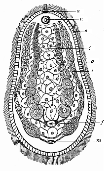 Fig. 239—Aphanostomum Langii (Haeckel), a primitive worm of the platodaria class, of the order of Cryptocoela or Acoela. This new species of the genus Aphanostomum, named after Professor Arnold Lang of Zurich, was found in September, 1899, at Ajaccio in Corsica (creeping between fucoidea). It is one-twelfth of an inch long, one-twenty-fifth of an inch broad, and violet in colour. a mouth, g auditory vesicle, e ectoderm, i entoderm, o ovaries, a spermaries, f female aperture, m male aperture.
 |
The Turbellaria, with which the similar Platodaria were formerly classed, differ materially from them in the more advanced structure of their organs, and especially in having a central nervous system (vertical brain) and excretory renal canals (nephridia); both originate from the ectoderm. But between the two germinal layers a mesoderm is developed, a soft mass of connective tissue, in which the organs are embedded. The Turbellaria are still represented by a number of different forms, in both fresh and sea-water. The oldest of these are the very rudimentary and tiny forms that are known as Rhabdocœla on account of the simple construction of their gut; they are, as a rule, less than a quarter of an inch long and of a simple oval or lancet shape (Fig. 240). The surface is covered with ciliated epithelium, a stratum of ectodermic cells. The digestive gut is still the simple primitive gut of the gastræa (d), with a single aperture that is both mouth and anus (m). There is, however, an invagination of the ectoderm at the mouth, which has given rise to a muscular pharynx (sd). It is noteworthy that the mouth of the Turbellaria (like the primitive mouth of the Gastræa) may, in this class, change its position considerably in the middle line of the ventral surface; sometimes it lies behind (Opisthostomum), sometimes in the middle (Mesostomum), sometimes in front (Prosostomum). This displacement of the mouth from front to rear is very interesting, because it corresponds to a phylogenetic displacement of the mouth. This probably occurred in the Platode ancestors of most (or all?) of the Cœlomaria; in these the permanent mouth (metastoma) lies at the fore end (oral pole), whereas the primitive mouth (prostoma) lay at the hind end of the bilateral body.
In most of the Turbellaria there is a narrow cavity, containing a number of secondary organs, between the two primary germinal layers, the outer or animal layer of which forms the epidermis and the inner vegetal layer the visceral epithelium. The earliest of these organs are the sexual organs; they are very variously constructed in the Platode-class; in the simplest case there are merely two pairs of gonads or sexual glands—a pair of testicles (Fig. 241 h)
[ 223 ]
and a pair of ovaries (e). They open externally, sometimes by a common aperture (Monogonopora), sometimes by separate ones, the female behind the male (Digonopora, Fig. 241). The sexual glands develop originally from the two promesoblasts or primitive mesodermic cells (Fig. 83 p). As these earliest mesodermic structures extended, and became spacious sexual pouches in the later descendants of the Platodes, probably the two cœlom-pouches were formed from them, the first trace of the real body-cavity of the higher Metazoa (Enterocœla).
The gonads are among the oldest organs, the few other organs that we find in the Platodes between the gut-wall and body-wall being later evolutionary products. One of the oldest and most important of these are the kidneys or nephridia, which remove unusable matter from the body (Fig. 240 nc). These urinary or excretory organs were originally enlarged skin-glands—a couple of canals that run the length of the body, and have a separate or common external aperture (nm). They often have a number of branches. These special excretory organs are not found in the other Cœlenteria (Gastræads, Sponges, Cnidaria) or the Cryptocœla. They are first met in the Turbellaria, and have been transmitted direct from these to the Vermalia, and from these to the higher stems.
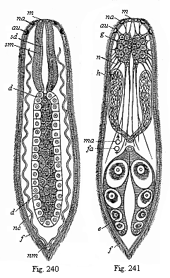 Fig. 240—A simple turbellarian (Rhabdocœlum). m mouth, sd gullet epithelium, sm gullet muscles, d gastric gut, nc renal canals, nm renal aperture, au eye, na olfactory pit. (Diagram.)
Fig. 241—The same, showing the other organs. g brain, au eye, na olfactory pit, n nerves, h testicles, ma male aperture, fa female aperture, e ovary, f ciliated epiderm. (Diagram.) |
Finally, there is a very important new organ in the Turbellaria, which we do not find in the Cryptocœla (Fig. 239) and their gastræad ancestors—the rudimentary nervous system. It consists of a couple of simple cerebral ganglia (Fig. 241 g) and fine nervous fibres that radiate from them; these are partly voluntary nerves (or motor fibres) that go to the thin muscular layer developing under the skin; and partly sensory nerves that proceed to the sense-cells of the ciliated epiderm (f). Many of the Turbellaria have also special sense-organs; a couple of ciliated smell pits (na), rudimentary eyes (au), and, less frequently, auditory vesicles.
On these principles I assume that the oldest and simplest Turbellaria arose from Platodaria, and these directly from bilateral Gastræads. The chief advances were the formation of gonads and nephridia, and of the rudimentary brain. On this hypothesis, which I advanced in 1872 in the first sketch of the gastræa-theory (Monograph on the Sponges), there is no direct affinity between the Platodes and the Cnidaria.
Next to the ancient stem-group of the Turbellaria come a number of more recent chordonia ancestors, which we class with the Vermalia or Helminthes, the unarticulated worms. These true worms (Vermes, lately also called Scolecida) are the difficulty or the lumber-room of the zoological classifier, because the various classes have very complicated relations to the lower Platodes on the one hand and the more advanced animals on the other. But if we exclude the Platodes and the Annelids from this stem, we find a fairly satisfactory unity of organisation
[ 224 ]
in the remaining classes. Among these worms we find some important forms that show considerable advance in organisation from the platode to the chordonia stage. Three of these phenomena are particularly instructive: (1) The formation of a true (secondary) body-cavity (cœloma); (2) the formation of a second aperture of the gut, the anus; and (3) the formation of a vascular system. The great majority of the Vermalia have these three features, and they are all wanting in the Platodes; in the rest of the worms at least one or two of them are developed.
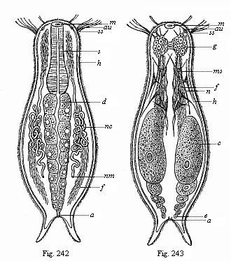 Figs. 242 and 243—Chætonotus, a rudimentary vermalian, of the group of Gastrotricha. m mouth, s gullet, d gut, a anus, g brain, n nerves, ss sensory hairs, au eye, ms muscular cells, h skin, f ciliated bands of the ventral surface, nc nephridia, nm their aperture, e ovaries.
 |
Next and very close to the Platodes we have the Ichthydina (Gastrotricha), little marine and fresh-water worms, about 1/250 to 1/1000 inch long. Zoologists differ as to their position in classification. In my opinion, they approach very close to the Rhabdocœla (Figs. 240, 241), and differ from them chiefly in the possession of an anus at the posterior end (Fig. 242 a). Further, the cilia that cover the whole surface of the Turbellaria are confined in the Gastrotricha to two ciliated bands (f) on the ventral surface of the oval body, the dorsal surface having bristles. Otherwise the organisation of the two classes is the same. In both the gut consists of a muscular gullet (s) and a glandular primitive gut (d). Over the gullet is a double brain (acroganglion, g). At the side of the gut are two serpentine prorenal canals (water-vessels or pronephridia, nc), which open on the ventral side (nm). Behind are a pair of simple sexual glands or gonads (Fig. 243 e).
While the Ichthydina are thus closely related to the Platodes, we have to go farther away for the two classes of Vermalia which we unite in the group of the “snout-worms” (Frontonia). These are the Nemertina and the Enteropneusta.
[ 225 ]
Both classes have a complete ciliary coat on the epidermis, a heritage from the Turbellaria and the Gastræads; also, both have two openings of the gut, the mouth and anus, like the Gastrotricha. But we find also an important organ that is wanting in the preceding forms—the vascular system. In their more advanced mesoderm we find a few contractile longitudinal canals which force the blood through the body by their contractions; these are the first blood-vessels.
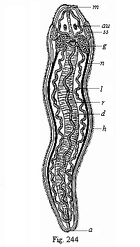 Fig. 244—A simple Nemertine. m mouth, d gut, a anus, g brain, n nerves, h ciliary coat, ss sensory pits (head-clefts), au eyes, r dorsal vessel, l lateral vessels. (Diagram.)
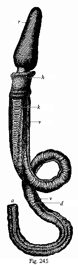
Fig. 245—A young Enteropneust (Balanaglossus). (From Alexander Agassiz.) r acorn-shaped snout, h neck, k gill-clefts and gill-arches of the fore-gut, in long rows on each side, d digestive hind-gut, filling the greater part of the body-cavity, v intestinal vein or ventral vessel, lying between the parallel folds of the skin, a anus. |
 |
[ 226 ]
The Nemertina were formerly classed with the much less advanced Turbellaria. But they differ essentially from them in having an anus and blood-vessels, and several other marks of higher organisation. They have generally long and narrow bodies, like a more or less flattened cord; there are, besides several small species, giant-forms with a width of 1/5 to 2/5 inch and a length of several yards (even ten to fifteen). Most of them live in the sea, but some in fresh water and moist earth. In their internal structure they approach the Turbellaria on the one hand and the higher Vermalia (especially the Enteropneusta) on the other. They have a good deal of interest as the lowest and oldest of all animals with blood. In them we find blood-vessels for the first time, distributing real blood through the body. The blood is red, and the red colouring-matter is hæmoglobin, connected with elliptic discoid blood-cells, as in the Vertebrates. Most of them have two or three parallel blood-canals, which run the whole length of the body, and are connected in front and behind by loops, and often by a number of ring-shaped pieces. The chief of these primitive blood-vessels is the one that lies above the gut in the middle line of the back (Fig. 244 r); it may be compared to either the dorsal vessel of the Articulates or the aorta of the Vertebrates. To the right and left are the two serpentine lateral vessels (Fig. 244 l).
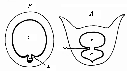 Fig. 246—Transverse section of the branchial gut. A of Balanoglossus, B of Ascidia. r branchial gut, n pharyngeal groove, * ventral folds between the two. Diagrammatic illustration from Gegenbaur, to show the relation of the dorsal branchial-gut cavity (r) to the pharyngeal or hypobranchial groove (n).
 |
After the Nemertina, I take (as distant relatives) the Enteropneusta; they may be classed together with them as Frontonia or Rhyncocœla (snout-worms). There is now only one genus of this class, with several species (Balanoglossus); but it is very remarkable, and may be regarded as the last survivor of an ancient and long-extinct class of Vermalia. They are related, on the one hand, to the Nemertina and their immediate ancestors, the Platodes, and to the lowest and oldest forms of the Chordonia on the other.
The Enteropneusta (Fig. 245) live in the sea sand, and are long worms of very simple shape, like the Nemertina. From the latter they have inherited: (1) The bilateral type, with incomplete segmentation; (2) the ciliary coat of the soft epidermis; (3) the double rows of gastric pouches, alternating with a single or double row of gonads; (4) separation of the sexes (the Platode ancestors were hermaphroditic); (5) the ventral mouth, underneath a protruding snout; (6) the anus terminating the simple gut-tube; and (7) several parallel blood-canals, running the length of the body, a dorsal and a ventral principal stem.
On the other hand, the Enteropneusta differ from their Nemertine ancestors in several features, some of which are important, that we may attribute to adaptation. The chief of these is the branchial gut (Fig. 245 k). The anterior section of the gut is converted into a respiratory organ, and pierced by two rows of gill-clefts; between these there is a branchial (gill) skeleton, formed of rods and plates of chitine. The water that enters at the mouth makes its exit by these clefts. They lie in the dorsal half of the fore-gut, and this is completely separated from the ventral half by two longitudinal folds (Fig. 246 A*). This ventral half, the glandular walls of which are clothed with ciliary epithelium and secrete mucus, corresponds to the pharyngeal or hypo-branchial groove of the Chordonia (Bn), the important organ from which the later thyroid gland is developed in the Craniota (cf. p. 184). The agreement in the structure of the branchial gut of the Enteropneusts, Tunicates, and Vertebrates was first recognised by Gegenbaur (1878); it is the more significant as at first we find only a couple of gill-clefts in the young animals of all three groups; the number gradually increases. We can infer from this the common descent of the three groups with all the more
[ 227 ]
confidence when we find the Balanoglossus approaching the Chordonia in other respects. Thus, for instance, the chief part of the central nervous system is a long dorsal neural string that runs above the gut and corresponds to the medullary tube of the Chordonia. Bateson believes he has detected a rudimentary chorda between the two.
Of all extant invertebrate animals the Enteropneusts come nearest to the Chordonia in virtue of these peculiar characters; hence we may regard them as the survivors of the ancient gut-breathing Vermalia from which the Chordonia also have descended. Again, of all the chorda-animals the Copelata (Fig. 225) and the tailed larvæ of the ascidia approach nearest to the young Balanoglossus. Both are, on the other hand, very closely related to the Amphioxus, the Primitive Vertebrate of which we have considered the importance (Chapters XVI and XVII). As we saw there, the unarticulated Tunicates and the articulated Vertebrates must be regarded as two independent stems, that have developed in divergent directions. But the common root of the two stems, the extinct group of the Prochordonia, must be sought in the vermalia stem; and of all the living Vermalia those we have considered give us the safest clue to their origin. It is true that the actual representatives of the important groups of the Copelata, Balanoglossi, Nemertina, Icthydina, etc., have more or less departed from the primitive model owing to adaptation to special environment. But we may just as confidently affirm that the main features of their organisation have been preserved by heredity.
We must grant, however, that in the whole stem-history of the Vertebrates the long stretch from the Gastræads and Platodes up to the oldest Chordonia remains by far the most obscure section. We might frame another hypothesis to raise the difficulty—namely, that there was a long series of very different and totally extinct forms between the Gastræa and the Chordæa. Even in this modified chordæa-theory the six fundamental organs of the chordula would retain their great value. The medullary tube would be originally a chemical sensory organ, a dorsal olfactory tube, taking in respiratory-water and food by the neuroporus in front and conveying them by the neurenteric canal into the primitive gut. This olfactory tube would afterwards become the nervous centre, while the expanding gonads (lying to right and left of the primitive mouth) would form the cœloma. The chorda may have been originally a digestive glandular groove in the dorsal middle line of the primitive gut. The two secondary gut-openings, mouth and anus, may have arisen in various ways by change of functions. In any case, we should ascribe the same high value to the chordula as we did before to the gastrula.
In order to explain more fully the chief stages in the advance of our race, I add the hypothetical sketch of man’s ancestry that I published in my Last Link [a translation by Dr. Gadow of the paper read at the International Zoological Congress at Cambridge in 1898]:—
[ 228 ]
A.—Man’s Genealogical Tree, First Half:
EARLIER SERIES OF ANCESTORS,
WITHOUT FOSSIL EVIDENCE.
| Chief Stages |
Ancestral Stem-groups |
Living Relatives of
Ancestors |
Stages 1–5:
Protist
ancestors
Unicellular
organisms.
1–2:
Prototypes
3–5:
Protozoa |
1. Monera
Without nucleus
2. Algaria
Unicellular algæ
|
1. Chromacea
(Chroococcus)
Phycochromacea
2. Paulotomea
Palmellacea
Eremosphæra |
3. Lobosa
Unicellular (amœbina)
rhizopods
4. Infusoria
Unicellular
5. Blastæades
Multicellular hollow spheres
|
3. Amœbina
Amœba Leucocyta
4. Flagellata
Euflagellata
Zoomonades
5. Catallacta
Magosphæra, Volvocina,
Blastula |
Stages 6–11:
Invertebrate
metazoa
ancestors
6–8:
Cœlenteria
without anus and
body-cavity
9–11:
Vermalia, with
anus and
body-cavity |
6. Gastræades
With two germ-layers
7 Platodes I
Platodaria
(without nephridia)
8. Platodes II
Platodinia (with nephridia) |
6. Gastrula
Hydra, Olynthus,
Gastremaria
7. Cryptocœla
Convoluta, Porporus
8. Rhabdocœla
Vortex, Monolus |
9. Provermalia
(Primitive worms)
Rotatoria
10. Frontonia
(Rhynchelminthes)
Snout-worms
11. Prochordonia
Chorda-worms
|
9. Gastrotricha
Trochozoa, Trochophora
10. Enteropneusta
Balanglossus
Cephalodiscus
11. Copelata
Appendicaria
Chordula-larvæ |
Stages 12–15:
Monorhina
ancestors
Oldest vertebrates
without jaws or
pairs of limbs,
single nose |
12. Acrania I
(Prospondylia)
13. Acrania II
More recent
14. Cyclostoma I
(Archicrania)
15. Cyclostoma II
More recent |
12. Amphioxus larvæ
13. Leptocardia
Amphioxus
14. Petromyzonta larvæ
15. Marsipobranchia
Petromyzonta |
B.—Man’s Genealogical Tree, Second Half:
LATER ANCESTORS, WITH FOSSIL EVIDENCE.
Geological
Periods |
Ancestral Stem-groups |
Living Relatives of
Ancestors |
| Silurian | 16. Selachii
Primitive fishes
Proselachii | 16. Natidanides
Chlamydoselachius
Heptanchus |
| Silurian | 17. Ganoids
Plated-fishes
Proganoids | 17. Accipenserides
(Sturgeons)
Polypterus |
| Devonian | 18. Dipneusta
Paladipneusta | 18. Neodipneusta
Ceratodus
Proptopterus |
| Carboniferous | 19. Amphibia
Stegocephala | 19. Phanerobranchia
Salamandrina
(Proteus, triton) |
| Permian | 20. Reptilia
Proreptilia | 20. Rhynchocephalia
Primitive lizards
Hatteria |
| Triassic | 21. Monotrema
Promammalia | 21. Ornithodelphia
Echidna
Ornithorhyncus |
| Jurassic | 22. Marsupialia
Prodidelphia | 22. Didelphia
Didelphys
Perameles |
| Cretaceous | 23. Mallotheria
Prochoriata | 23. Insectivora
Erinaceida
(Ictopsia +) |
| Older Eocene | 24. Lemuravida
Older lemurs
Dentition 3. 1. 4. 3. | 24. Pachylemures
(Hyopsodus +)
(Adapis +) |
| Neo-Eocene | 25. Lemurogona
Later lemurs
Dentition 2. 1. 4. 3. | 25. Autolemures
Eulemur
Stenops |
| Oligocene | 26. Dysmopitheca
Western apes
Dentition 2. 1. 3. 3. | 26. Platyrrhinæ
(Anthropops +)
(Homunculus +) |
| Older Miocene | 27. Cynopitheca
Dog-faced apes (tailed) | 27. Papiomorpha
Cynocephalus |
| Neo-Miocene | 28. Anthropoides
Man-like apes (tail-less) | 28. Hylobatida
Hylobates
Satyrus |
| Pliocene | 29. Pithecanthropi
Ape-men (alali, speechless) | 29. Anthropitheca
Chimpanzee
Gorilla |
| Pleistocene | 30. Homines
Men with speech | 30. Weddahs
Australian negroes |
Title and Contents
Vol. II Title and Contents
Glossary
Chapter XIX
Chapter XXI
Figs. 1–209
Figs. 210–408